Human Anatomy Female Abdomen : Human Anatomy Abdomen Female Anatomy Drawing Diagram - They are separated by frank h.. National library of medicine was used as the basis to build an. Posted on april 11, 2019. This hd wallpaper anatomy of female abdomen has viewed by 846 users. There are many pics about muscles human body diagram out there. Human anatomy lesson 15 abdomen.
They are separated by frank h. • we're going to take apart a plastic anatomy model and see what we. Both pass through a skeletal muscle (voluntary) sphincter in the urogenital. If you want to learn how to read ct scans of the abdomen and pelvis proficiently, this video is an excellent starting point. Data set from the u.s.
Female Abdomen Youtube from i.ytimg.com The standard position of the uterus is anteverted and anteflexed. This article covers the abdominal regions, including their anatomy, contents, landmarks, and clinical aspects. The abdomen is the largest cavity in the body. It is of an oval shape, the extremities of the oval being directed upward and downward. This full color custom medical exhibit features an anterior and sagittal view of the normal anatomy of the female reproductive system, an enlarged anterior view of the left fallopian tube and ovary is included. Of human anatomy and different types of motion, inspiring more realistic and energetic figurative art. Data set from the u.s. Image from marieb et al., human anatomy, 7th edition, pearson education, 2014.
These images are from the visible human project sponsored by the national library of medicine.
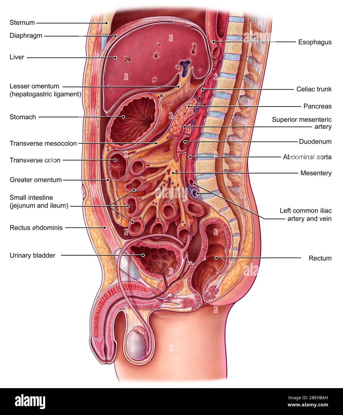
The human abdomen is that part in the front of our body between the chest and the waist line. Thus, the right side of the image is the patient's left. This full color custom medical exhibit features an anterior and sagittal view of the normal anatomy of the female reproductive system, an enlarged anterior view of the left fallopian tube and ovary is included. The bones of the abdomen are made up of the lumbar. Human female (vhf) for laparoscopic surgery training. The four anatomical regions of the abdomen are known as quadrants. Blood vessels, lymphatic drainage and nerves of the abdomen. Diseases affecting any of these organs could result in abdominal pain. If you want to learn how to read ct scans of the abdomen and pelvis proficiently, this video is an excellent starting point. The liver, stomach, large intestines, rectum, uterus, vaginal. Female anatomy, early 17th c wellcome l0011866.jpg 1,178 × 1,707; These images are arranged in radiographic view, as though you were looking up from the patient's feet toward the head. A regional study of human structure.
National library of medicine was used as the basis to build an. Female abdominal thoracic anatomy medical illustration. It is of an oval shape, the extremities of the oval being directed upward and downward. Organs shown and labeled are: These images are from the visible human project sponsored by the national library of medicine.
Front View Of Female Chest And Abdominal Muscles Anatomy In Pink X Ray Outline Full Color 3d Computer Generated Illustration On Black Background Stock Photo 1428r 1422 Superstock from cdn.superstock.com Two ways of dividing the abdomen into regions. Anatomy of liver the liver is a reddish brown organ with four lobes of unequal size and shape. Organs shown and labeled are: It is of an oval shape, the extremities of the oval being directed upward and downward. its musculomembranous walls surround a large cavity (the 28. The human abdomen is that part in the front of our body between the chest and the waist line. Human body anatomy yoga anatomy human anatomy and physiology muscle anatomy knee muscles anatomy anatomy study. The video covers the most.
To delineate organ outlines and.
They are separated by frank h. its musculomembranous walls surround a large cavity (the 28. Diseases affecting any of these organs could result in abdominal pain. If you want to learn how to read ct scans of the abdomen and pelvis proficiently, this video is an excellent starting point. Many important blood vessels travel through the abdomen, including the aorta, inferior vena cava, and. Human female (vhf) for laparoscopic surgery training. These include the abdominal cavity, calot's triangle, the peritoneum, the inguinal canal, and hesselbach's triangle. Organs shown and labeled are: The human abdomen is that part in the front of our body between the chest and the waist line. Two ways of dividing the abdomen into regions. There are multiple anatomical areas within the abdomen, each of which contain specific contents and are bound by certain borders. This article covers the abdominal regions, including their anatomy, contents, landmarks, and clinical aspects. The liver, stomach, large intestines, rectum, uterus, vaginal.
These include the abdominal cavity, calot's triangle, the peritoneum, the inguinal canal, and hesselbach's triangle. Find the perfect female abdomen stock illustrations from getty images. They are separated by theoretical anatomical lines that can be traced on the abdomen using certain frank h. The female and male urethrae. • we're going to take apart a plastic anatomy model and see what we.
Abdominal Organs Illustration Stock Photo Alamy from c8.alamy.com Image from marieb et al., human anatomy, 7th edition, pearson education, 2014. Anatomy of liver the liver is a reddish brown organ with four lobes of unequal size and shape. Diseases affecting any of these organs could result in abdominal pain. We think this is the most useful anatomy. Don't forget to share this picture with others via facebook, twitter, pinterest or other social medias! The four anatomical regions of the abdomen are known as quadrants. The bones of the abdomen are made up of the lumbar. Both pass through a skeletal muscle (voluntary) sphincter in the urogenital.
Human female (vhf) for laparoscopic surgery training.
Don't forget to share this picture with others via facebook, twitter, pinterest or other social medias! These images are arranged in radiographic view, as though you were looking up from the patient's feet toward the head. It is of an oval shape, the extremities of the oval being directed upward and downward. Female and male anatomy sigmoid colon, sigmoid mesocolon, rectosigmoid junction, peritoneal reflection, rectovesical pouch. The video covers the most. National library of medicine was used as the basis to build an. The abdomen (colloquially called the belly, tummy, midriff or stomach) is the part of the body between the thorax (chest) and pelvis, in humans and in other vertebrates. We think this is the most useful anatomy. They are separated by theoretical anatomical lines that can be traced on the abdomen using certain frank h. There are many pics about muscles human body diagram out there. Diseases affecting any of these organs could result in abdominal pain. The four anatomical regions of the abdomen are known as quadrants. Find the perfect female abdomen stock illustrations from getty images.


0 Komentar